Fig. 8.
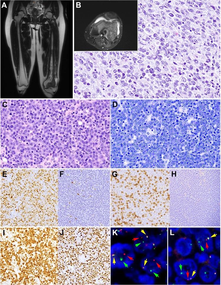
Primary diffuse large B-cell lymphoma of the bone (A–B). Case LYWS-1231 courtesy of Y-Ch. Liu. A–B Imaging studies (MRI) of the left femur shows a lobulated mass extended from the mid diaphysis into the distal epiphysis including both the medial and lateral femoral condyles. B The bone biopsy showed a polymorphic infiltrate predominantly of large cells confirming the diagnosis of diffuse large B-cell lymphoma. C–L Diffuse large B-cell lymphoma with CCND1 rearrangement. Case LYWS-1380 courtesy of K.S. Kurz. C Lymph node biopsy with effaced architecture by a diffuse infiltrate of predominantly medium-sized to large blast cells. D Giemsa stain reveals that the tumor cells have basophilic cytoplasm and round to oval nuclei with open chromatin and prominent nucleolus. E Cyclin D1 is positive in the tumor cells. F In other areas of the lymph node the tumor cells were cyclin D1 negative. G CD5 stain is positive in the reactive T cells but negative in the tumor cells. H SOX11 remains negative. I MUM1 is positive in the majority of tumor cells whereas CD10 remained negative (not shown). J Ki-67 demonstrate a high proliferation rate. K–L Interphase FISH analysis using break apart probes for CCND1 and MYC reveals 1 fusion signal (yellow arrow), one red signal (red arrow), and one green signal (green arrow) indicative of CCND1 (K) and MYC (L) rearrangements

