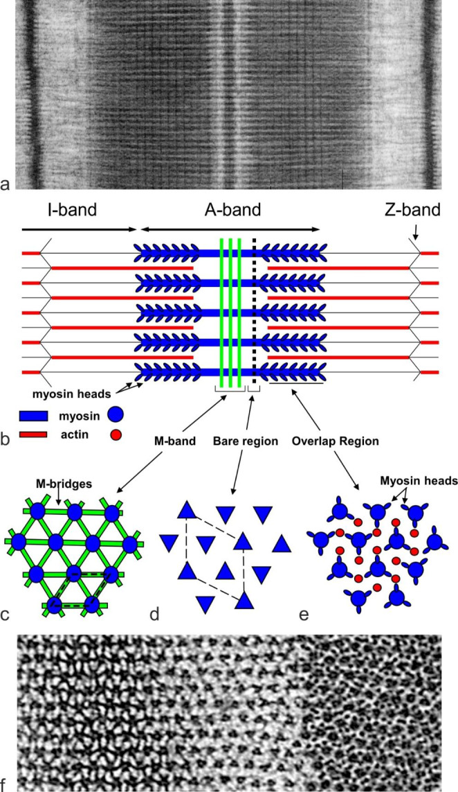
Fig. 1.

Vertebrate striated muscle sarcomere and the myosin filament superlattice. (a) Electron micrograph of frog sartorius muscle sarcomere and (b) schematic diagram of the sarcomere. The sarcomere is bounded by the Z-bands from which emanate actin filaments which overlap with the myosin filaments in the A-band. Contraction of muscle occurs when crossbridges on the myosin filaments interact with actin filaments to bring them closer to the centre of the A-band. The bipolar myosin filaments are crosslinked at the centre by the M-band assembly (b,c) which determines their relative rotation about the long axis. (c,d,e) Show schematic views of cross-sections in the M-band, bare region and the crossbridge region, respectively. (f) Shows electron micrographs of transverse sections through the M-band (left), bare region (centre) and crossbridge region (right). In the crossbridge region, the filament cross-section profile is indistinct hence the axial rotations of the filaments cannot be determined. In the bare region between the A-band and the start of the crossbridge region, the myosin filaments have a triangular profile (d and f centre) and this enables determination of the axial rotations of the filaments within 60o. In a hexagonal lattice we can define a simple unit cell (c, dashed outline). In certain vertebrate muscles, the myosin filaments tend to have identical orientations for second-next nearest neighbours (d) and so are arranged on a superlattice (dashed outline). Parts b,c,d,e adapted from Millane et al. (2021)
