
Fig. 3.
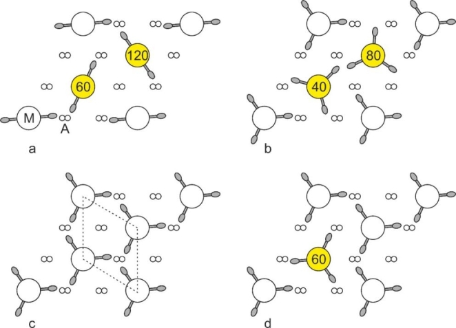
Crossbridge environments within a thin slice of vertebrate striated muscle within a single superlattice unit cell. M and A depict myosin and actin filaments, respectively. By definition, the rotations of the corner filaments of the superlattice unit cell are identical. Where the rotations are different to the corner rotations, they are coloured yellow. Initially, perfect superlattice arrangements were proposed (a and b). (a) For a 2-stranded myosin filament (where rotations of 0o and 180o are equivalent), Huxley and Brown (1967) proposed 60o rotations between neighbouring filaments. (b) For a 3-stranded myosin filament, Squire suggested 40o rotations between neighbouring filaments (Squire 1974). Perfect superlattice arrangements were not found however in electron micrographs, but the arrangement is a statistical superlattice (Luther and Squire 1980). Crossbridge arrangements for a simple lattice (c) and a superlattice muscle (d). (c) In simple lattice muscle (unit cell shown in dashed lines), all myosin filament rotations are identical and the actin filaments are approached by three or no crossbridges. In superlattice muscles (d), the two internal filaments are arranged statistically and the actin filaments are approached by one or two crossbridges