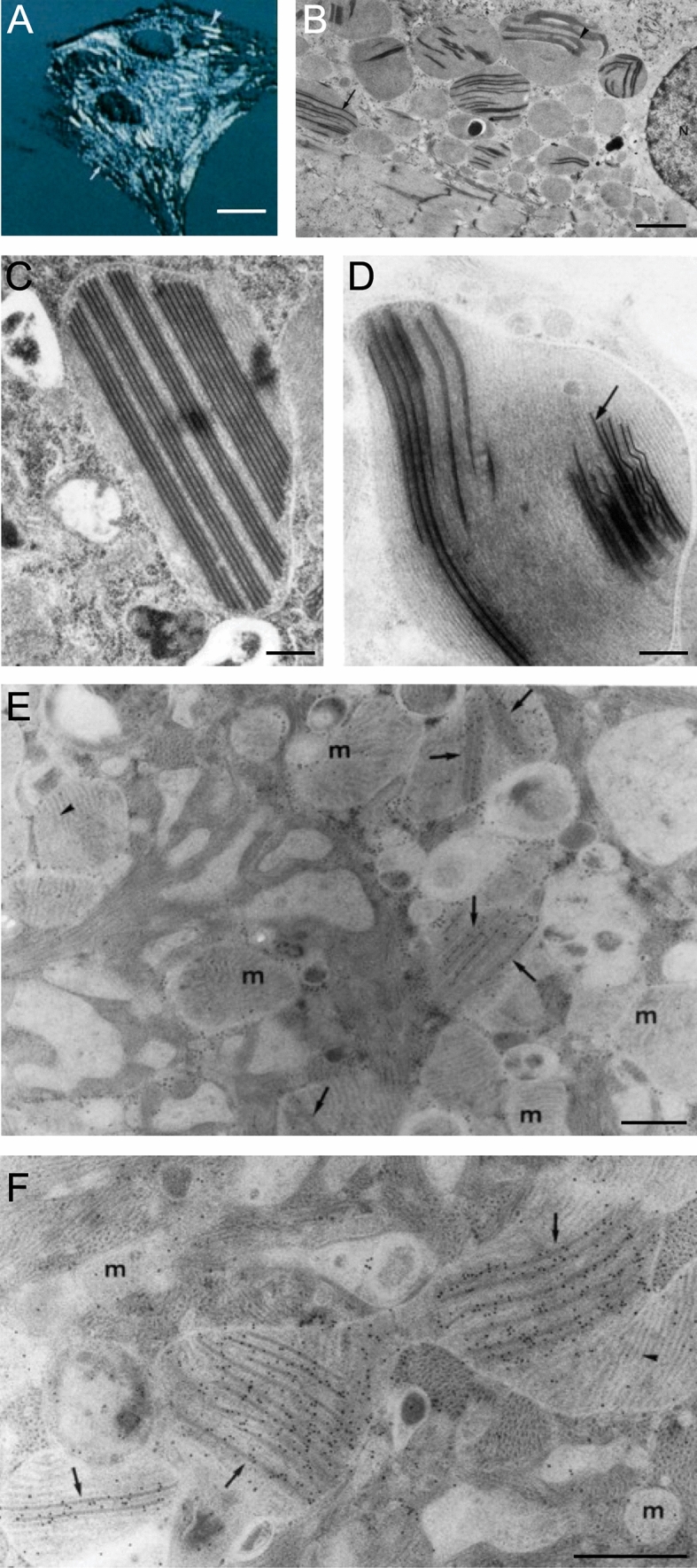
Fig. 4.

Cultured adult rat ventricular cardiomyocyte (ARVC). A Artificially shadowed confocal microscopy image showing giant cylindrical shaped mitochondria (arrowhead). B Corresponding low magnification transmission electron microscopy image shows the presence of electron-dense inclusions in giant mitochondria. The inclusions can appear as parallel straight lines (arrow) or sheet liked wavy structures (arrowhead). N—nucleus. Periodicity was identified between the parallel straight lines C and within the linear structures (D, arrow). E–F ARVCs gold-labelled with mitochondrial creatine kinase antibody are localized at the site of both forms of the mitochondrial inclusions, but largely absent from mitochondria (m) without inclusions. Scale bars, 15 μm (A); 3 μm (B); 0.45 μm (C & D); 0.5 μm (E & F). (Figs. 1F, 3 and 4: Journal of Cell Biology by The Rockefeller University Press [16]. Reproduced and lettering modified with permission of The Rockefeller University Press in the format Journal/Magazine via Copyright Clearance Center)
