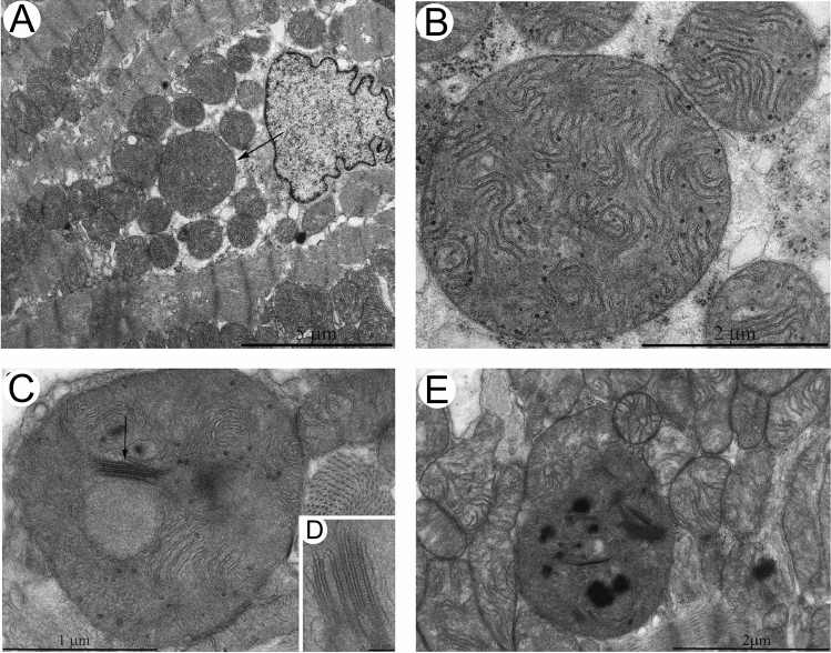
Fig. 5.
Electron microscopy observations of mitochondria in the naked mole rat. A shows the presence of a large mitochondrion (arrow) at 5 years of age. Note, the high number of normal-sized mitochondria surrounding the enlarged mitochondrion. B High magnification of the giant mitochondrion from A which illustrates packed wave-like cristae. Note the electron-dense granules scattered throughout the mitochondrial matrix. C At 11-year-old, the large mitochondria show inclusion of highly ordered cristae bundles and a clear reduction in cristae packing. D High magnification of the cristae bundles in C which are arranged in parallel, with a track-like appearance between the double-membraned structures termed membrane junctions or intra-crystal junctions. E Overview of the disrupted mitochondria ultrastructure in 11 year-old naked mole rats with possible sheet-like inclusions. Scale bars, 5 μm (A); 2 μm (B); 1 μm (C); 0.1 μm (D); 2 μm (E). (Figs. 5 and 8: International Journal of Molecular Sciences by MDPI [5]. Reproduced and lettering modified with permission of MDPI in the format Journal/Magazine via MDPI Open Access Policy)

