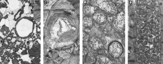
Fig. 7.
Electron microscopy micrographs showing ultrastructural changes of mitochondria in patients with mtDNA mutations. A Ring-shaped mitochondria. B Giant mitochondria with membrane fusion and circular cristae. C Concentric cristae. D Giant ‘organelles’ containing irregularly whorled and undulated cristae. End magnifications, × 5600 (A); × 16,000 (B); × 9600 (C); × 9100 (D). (Fig. 1: American Journal of Pathology by Elsevier [4]. Reproduced and lettering modified with permission of Elsevier’s Open Access Content License policy for the American Society for Investigative Pathology in the format Journal/Magazine and subject to proper acknowledgement of the original source)

