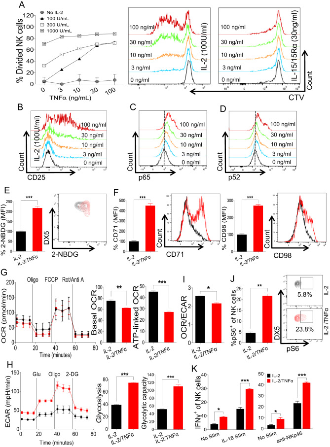
Fig. 3.
NK cell proliferation and metabolic activity under stimulation with TNFα ex vivo. A NK cells were enriched from the spleens of naive C57BL/6 mice and stained with cell trace violet dye followed by stimulation with the indicated doses of rhIL-2, IL-15/IL-15Rα complex, and TNFα ex vivo for 3 days, and the percentage of dividing NK cells was analyzed. B CD25 upregulation on NK cells following stimulation with the indicated doses of IL-2 and TNFα. C–F NK cells were enriched from the spleens of naive C57BL/6 mice and stimulated with TNFα ex vivo for 48 h. 100 U/ml rhIL-2 was added to maintain NK cell survival. Representative plots depict the expression of (C) pNFkB-p65 and (D) pNFkB-p52. The graphs represent (E) glucose uptake by NK cells as measured by the MFI of 2-NBDG and (F) the expression of CD71 and CD98 in cytokine-stimulated NK cells. G–I Freshly isolated NK cells were flow-sorted followed by ex vivo TNFα stimulation for 48 h, and the oxygen consumption rate (OCR) and extracellular acidification rate (ECAR) were analyzed. J Cells were prepared as in (C), and the proportion of pS6+ cells among the total NK cell population was calculated. K NK cells were enriched from the spleens of naive C57BL/6 mice and treated with TNFα ex vivo for 18 h in the presence of 100 U/ml rhIL-2 followed by stimulation with plate-coated anti-NKp46 or IL-18 for an additional 5 h, and the proportion of IFNγ+ cells among the total NK cell population was measured. The MFI expression is presented as a percentage relative to the MFI of control cells as 100. Data are from one experiment representative of three independent experiments, with at least two replicates per group. Data represent the mean ± SD. *p < 0.05; **p < 0.01; ***p < 0.001

