Fig. 1.
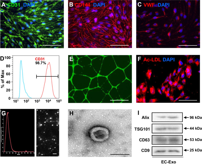
Characterization of hiPSC-ECs and exosomes secreted from hiPSC-ECs. A–C hiPSCs were differentiated into endothelial cells (hiPSC-ECs). hiPSC-ECs were characterized via immunofluorescent analyses of the expressions of A CD31, B CD144, and C von Willebrand factor (VWF). Nuclei were counterstained with 4′,6-diamidino-2-phenylindole (DAPI) (bar = 100 μm). D The purity of hiPSC-ECs was determined by FACS analysis of CD31. E hiPSC-ECs were suspended in a culture medium supplemented with 50 ng/mL vascular endothelial growth factor (VEGF) and then plated on Matrigel; 24 h later, the cells were labeled with calcein, and tube formation was evaluated under a light microscope (bar = 100 μm). F The uptake of Dil-conjugated acetylated low-density lipoprotein (Ac-LDL) was evaluated in hiPSC-ECs; nuclei were counterstained with DAPI (bar = 100 μm). G–I Exosomes were isolated from the culture medium of hiPSC-ECs; then, G exosome size was evaluated via nanoparticle tracking analysis (bar = 500 nm), H exosome morphology was evaluated via electron microscopy (bar = 100 nm), and I the presence of exosome marker proteins (ALG-2-interacting protein X [Alix], tumor susceptibility gene 101 protein [TSG101], CD63, and CD9) was evaluated via Western blot. Full-length blots are presented in Additional file 1: Fig. S1

