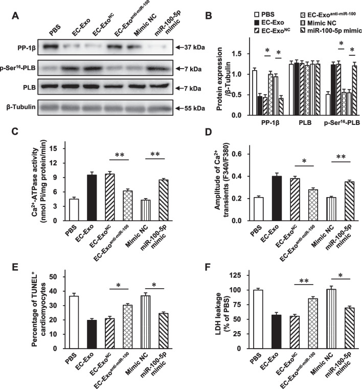
Fig. 8.
miR-100-5p loss abolishes, while miR-100-5p mimic reproduces, the part of protection of hiPSC-EC exosomes on cardiomyocytes against OGD injury. hiPSC-CMs were separately treated with PBS, mimic negative control (NC), miR-100-5p mimic, EC-Exo, EC-ExoNC, and EC-Exoanti−miR−100−5p under OGD conditions. A–B Protein samples were collected from the above-mentioned groups, and Western blot was performed to assess the protein expression levels of PP-1β, p-Ser16-PLB, and PLB. A Representative immunoblots. Full-length blots are presented in Additional file 1: Fig. S10. B Quantitative analysis of protein expression levels. C SERCA activity was assessed using a Ca2+-pump ATPase enzyme assay kit. D hiPSC-CMs were incubated with a Ca2+ indicator (Fura-2 AM) and stimulated at 0.5 Hz; then, Ca2+ transients were recorded and quantified. E The activity of LDH released in the culture media was measured. F TUNEL+ cardiomyocytes were assessed. Quantitative data are presented as mean ± SEM. n = 4 independent experiments in B and n = 5 independent experiments in C–F. Significance was evaluated via one-way ANOVA followed by Tukey’s post hoc test in (B–F). *p < 0.05 and **p < 0.01

