Fig. 3.
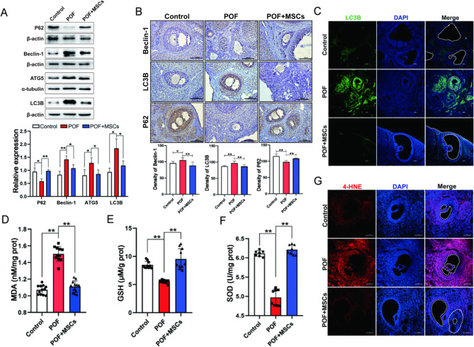
The oxidative damage and excessive autophagy in ovarian GCs were alleviated via hC-MSCs. A The Western blotting images and quantitation of autophagy associated proteins expression in ovaries. B The immunohistochemistry images and quantitation of autophagy-associated proteins expression in ovaries; scale bar: 100 μm. C The immunofluorescence images of LC3B expression in ovaries; scale bar: 100 μm. MDA (D), GSH (E), and SOD activity (F) levels in ovaries. G The lipid peroxidation producer 4-HNE in ovaries; scale bar: 100 μm. *p < 0.05. **p < 0.01

