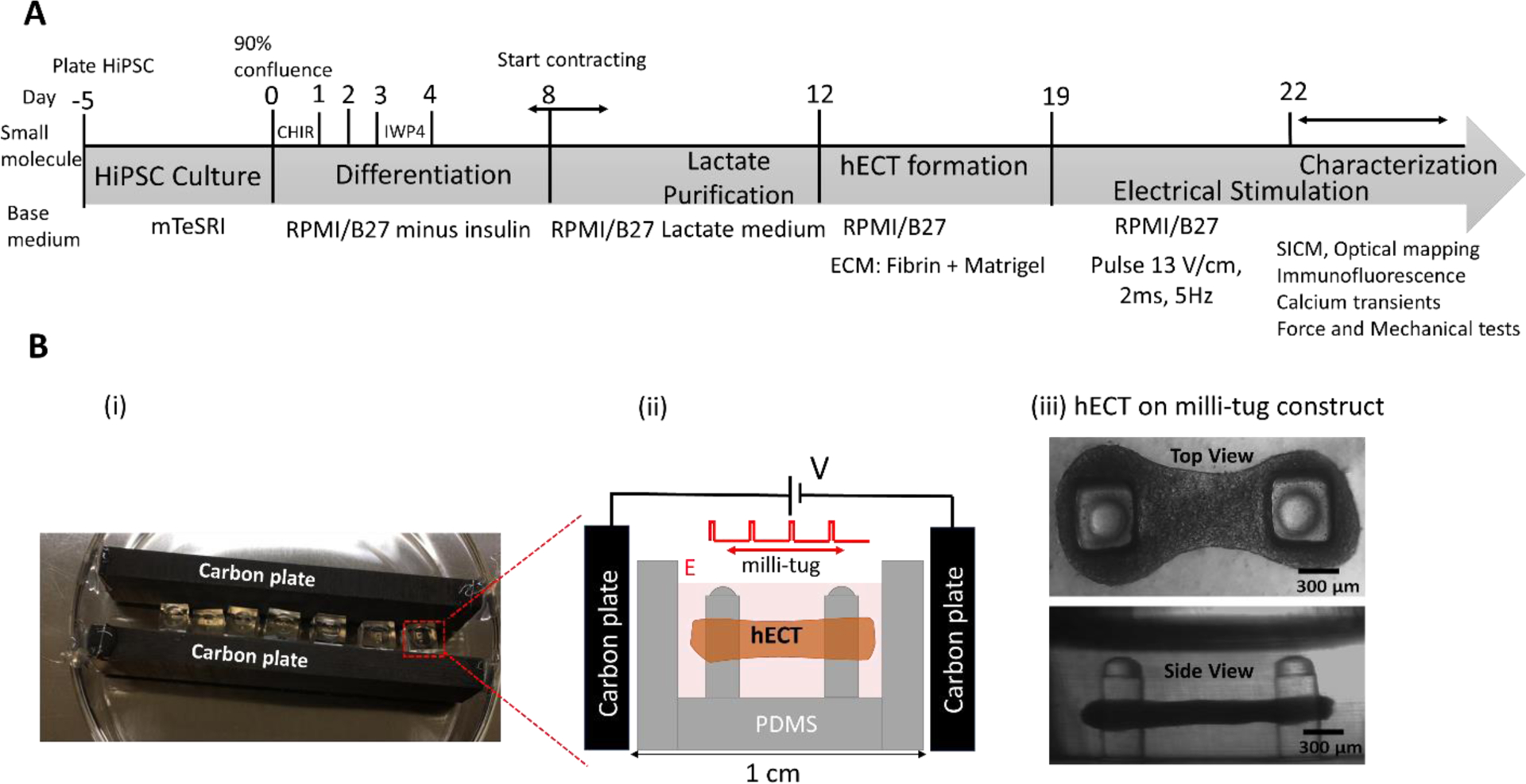
Figure 1.

(A) Timeline for the formation of hiPSC-CMs derived hECT and the following characterizations. (B) (i) The photo image of the ES setup. (ii) The diagram illustrates the capacitive coupling configuration and the hECT in the milli-tug construct. (iii) The transmitted-light optical microscope images (both top and side views) of a live hECT in the milli-tug construct. Tissues were placed under ES on the first day of spontaneous beating.
