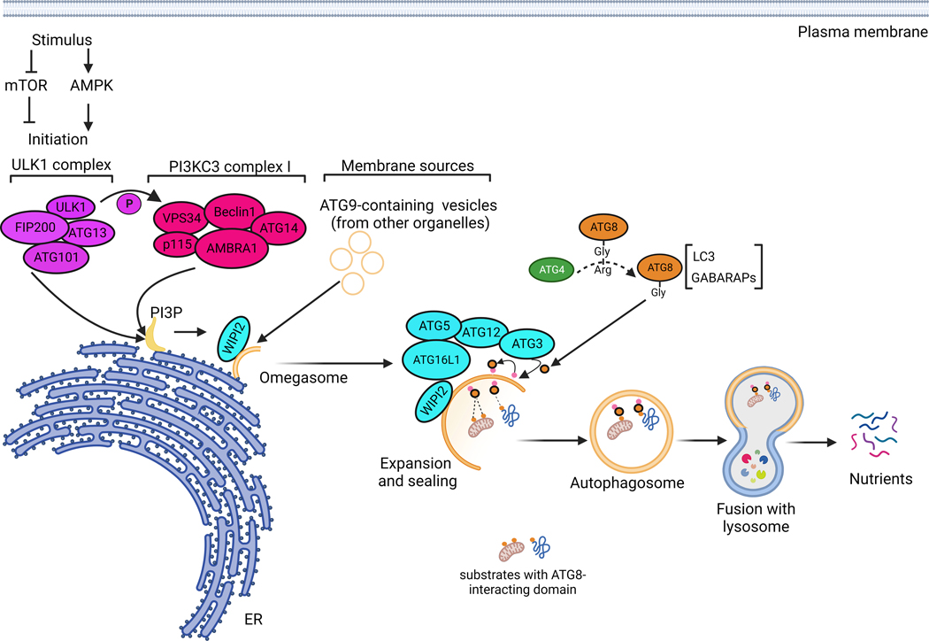
Figure 1. Molecular mechanisms of autophagy.
In response to environmental stresses, autophagy initiation is thought to take place at the endoplasmic reticulum membranes, where the ULK1 complex activates the PI3KC3 complex by phosphorylation. The PI3KC3 complex initiates the production of phosphatidylinositol-3-phosphate (PI3P) on ER subdomains forming a structure called the omegasome. In addition to PI3P provided by PI3KC3 complex, ATG9-containing small vesicles, originating from the membranes of other organelles, are involved in autophagy initiation. From the omegasome structure, a series of reactions involving a large panel of ATGs will take place to allow autophagosome elongation, sealing, maturation and fusion with lysosomes. ATG4, including ATG4B, cleaves Pro-ATG8 forms, such as LC3 and GABARAPL, to allow their conjugation into major phospholipids on the forming autophagosome. This lipidation reaction is catalysed by a complex involving WIPI2, ATG16L1, ATG5, ATG12, ATG7 and ATG3. The lipidation of ATG8s, such as LC3, is important for autophagosome elongation and maturation, but also for the interaction with cargos destined for autophagic degradation; it is therefore a major and critical step in the autophagic process.

