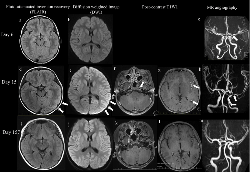
Figure 1.

Imaging findings. (a – c): on the 6th day after symptom onset, MRA of the head revealed narrowing of the left middle cerebral artery (c, arrow). Diffusion-weighted images (DWI) and fluid-attenuated inversion recovery (FLAIR) show no lesions. (d,e): on the 15th day after symptom onset, DWI and FLAIR show cortical high-intensity areas in the left cerebral hemisphere (arrow). (f,g): contrast-enhanced effects can be observed in the vessel walls of the left middle cerebral arteries, some of which are nodule-like or concentric (arrow). (h): MRA shows stenosis in the left internal carotid artery and left middle cerebral artery (arrow), and peripheral delineation is slightly poor on the affected side. (i – m): on the 157th day, after symptom onset abnormal findings are noted to have improved.
