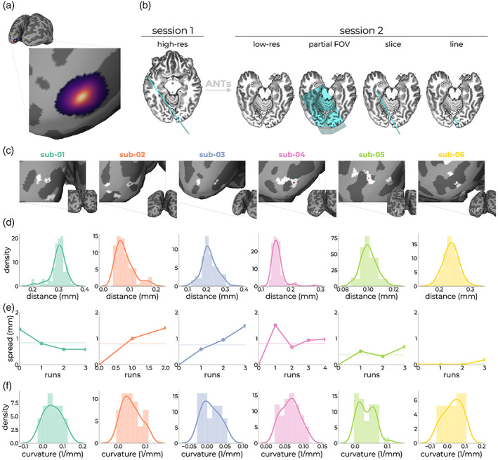
FIGURE 3.

Assessment of line‐placement accuracy using anatomical measures. The registration cascade from target vertex (a) to line‐scanning acquisition (b; outer right panel) is known after registering the anatomical image from Session 1 (high‐res) to the anatomical image from Session 2 (low‐res). Within Session 2, we acquired a partial field‐of‐view image (partial FOV), as well as the anatomical slice without OVS pulses (slice). From this slice, we created an image representing the nominal line (line). For each subject, we projected this nominal line image back to the surface from which the target vertex originated, showing sufficient overlap between the target vertex (red dot) and nominal line image (white patches) (c). The patches represent the location at which the nominal line image intersects with GM and looks scattered due to unfolding of the cortex. (d) Variation in registration outcomes after registration anatomies from Sessions 1 and 2, a hundred times for each subject. (e) Effect of subject motion by means of manual alignment of the single slice images on positional stability of the target coordinate. (f) Curvature distributions within the gray patch intersecting with the target location of (c). The curvature is measured as 1/r, where r is the radius of an inscribed circle. Since mean curvature is the average of the two principal curvatures, it has the units of 1/mm (https://surfer.nmr.mgh.harvard.edu/fswiki/MeanCurvature).
