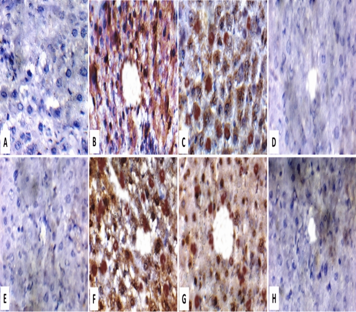
Figure 10.
Immunohistochemical staining of liver sections showing A] negative results for NF-κB in healthy control GI and vehicle group, B] marked positive results (++++) for NF-κB in injured untreated control GI, C] moderate positive result (+++) for NF-κB in injured free Mel (5 and 10 mg/kg) treated GII and GIII, D] negative results for NF-κB in injured Mel-PLGA NPs (5 and 10 mg/kg) treated GIV and GV, E] negative results for CRP in healthy control GI and vehicle group, F] marked positive results (++++) for CRP in injured untreated control GI, G] moderate positive result (+++) for CRP in injured free Mel (5 and 10 mg/kg) treated GII and GIII, H] negative results for CRP in injured Mel-PLGA NPs (5 and 10 mg/kg) treated GIV and GV.

