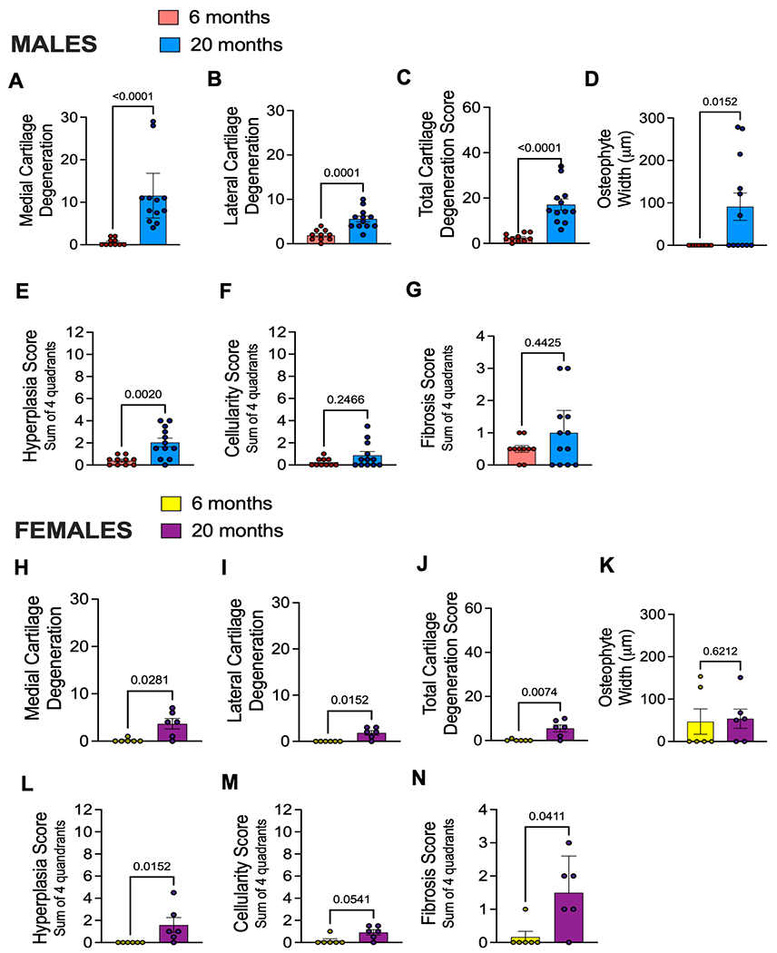
Fig. 1.

Aging is associated with mild knee osteoarthritis in mice. (A) Medial cartilage degeneration scores for male mice right knees aged 6 months (n=10; Mean ± SEM = 0.600 ± 0.267) or 20 months (n=12; Mean ± SEM = 11.5 ± 2.39) old as determined by OARSI scoring methods; (B) lateral cartilage degeneration for males aged 6 months (n=10; Mean ± SEM = 1.9 ± 0.3786) or 20 months (n=12; Mean ± SEM = 5.58 ± 0.656) ; (C) Total cartilage degeneration score for males aged 6 months (n=10; Mean ± SEM = 2.5 ± 0.543 or 20 months (n=12; Mean ± SEM = 17.13 ± 2.49) plotted as sum of medial and lateral compartments; (D) Osteophyte width for males plotted in micron size found in either medial or lateral compartments of the knee; (E) Synovitis hyperplasia score for males showing an average of two blinded scores (maximum score is 12); (F) Synovitis cellularity score for males showing an average of two blinded scores (maximum score is 12); (G) Synovitis fibrosis score for males showing an average of two blinded scores (maximum score is 4); (H – N) Same as in (A – G) but shown for female mice aged 6 months (n=6; Medial Mean ± SEM = 0.1667 ± 0.1667; Lateral Mean ± SEM = 0.0 ± 0.0; Total Mean ± SEM = 0.1667 ± 0.1667) or 20 months old (n=6; Medial Mean ± SEM = 3.667 ± 1.116; Lateral Mean ± SEM = 1.884 ± 0.4773; Total Mean ± SEM = 5.50 ± 1.586). Statistical analysis by Mann-Whitney test. Significant if p < 0.05. Error bars show Mean ± SEM.
