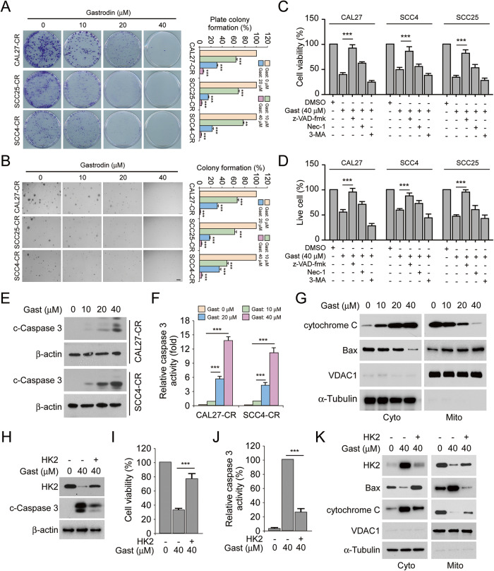
Fig. 4. Gastrodin induces endogenous apoptosis and decreases the protein level of HK2.
A Colony formation of CAL27-CR, SCC25-CR, and SCC4-CR cells treated with various concentrations gastrodin were measured by plate colony formation assay. **p < 0.01, ***p < 0.001. B Anchorage-independent growth of CAL27-CR, SCC25-CR, and SCC4-CR cells treated with various concentrations gastrodin were detected using soft agar assay. ***p < 0.001. C The viability of OSCC cells following various inhibitors and gastrodin (40 µM) treatment was evaluated using MTS analysis. ***p < 0.001. D The number of live cells in various inhibitors- and gastrodin-treated (40 µM) OSCC cells were measured using trypan blue exclusion analysis. ***p < 0.001. The protein levels of cleaved-caspase 3 (E) and caspase 3 activity (F) in CAL27-CR and SCC4-CR cells after treatment with various doses gastrodin. ***p < 0.001. G Isolating subcellular fractions of CAL27-CR cells treated with various doses gastrodin for IB assay. HK2 was transfected into CAL27-CR cells and the cells were treated with gastrodin (40 µM) for 24 h, followed by IB analysis (H), MTS analysis (I) and Caspase 3 activity assay (J). ***p < 0.001. K CAL27-CR cells were treated with gastrodin for 24 h following transfecting HK2. Separating subcellular fractions for IB assay.

