Fig. 1.
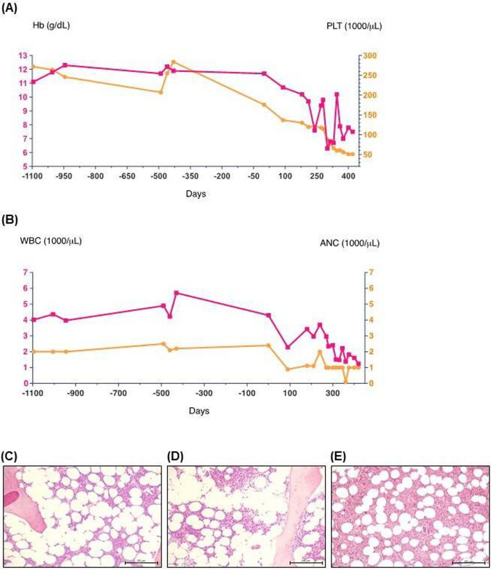
Case-based approach to aplastic anemia. A Timeline (days) of peripheral blood (PB) laboratory values, identified pancytopenia with a hemoglobin (Hb, purple) level of 7.6 g/dL (reference range 12–16 g/dL) without signs of hemolysis and a platelet (PLT, yellow) count of 51,000/µL (reference range 140,000–430,000 PLTs/µL). B Timeline (days) of a white blood cell (WBC, purple) count showed values of 1,240 cells/µL (reference range 4,500–11,000 cells/µL) and an absolute neutrophil (ANC, yellow) count of 1,000 cells/µL (count nadir reported, reference range 1,800–7,700 cells/µL). C–E Representative images of immunohistochemical staining in formalin-fixed 4 µm BM sections from the iliac crest. C Stat1 GOF variant patient, D idiopathic aplastic anemia and E non-aplastic benign anemia were stained with Haematoxilin-Eosin (DAKO, Golstrup, Denmark). Representative images showed a ratio of hematopoietic marrow to adipose tissue of 1:3 in our patient with STAT1 GOF variant (hypocellular condition to the patient's age). The erythroid and myeloid series markedly decreased, and a prevalence of more mature precursors was revealed. Sections were examined using an Olympus microscope (Olympus Italia, Rozzano, Italy). Original magnification × 10; scale bar = 100 µm

