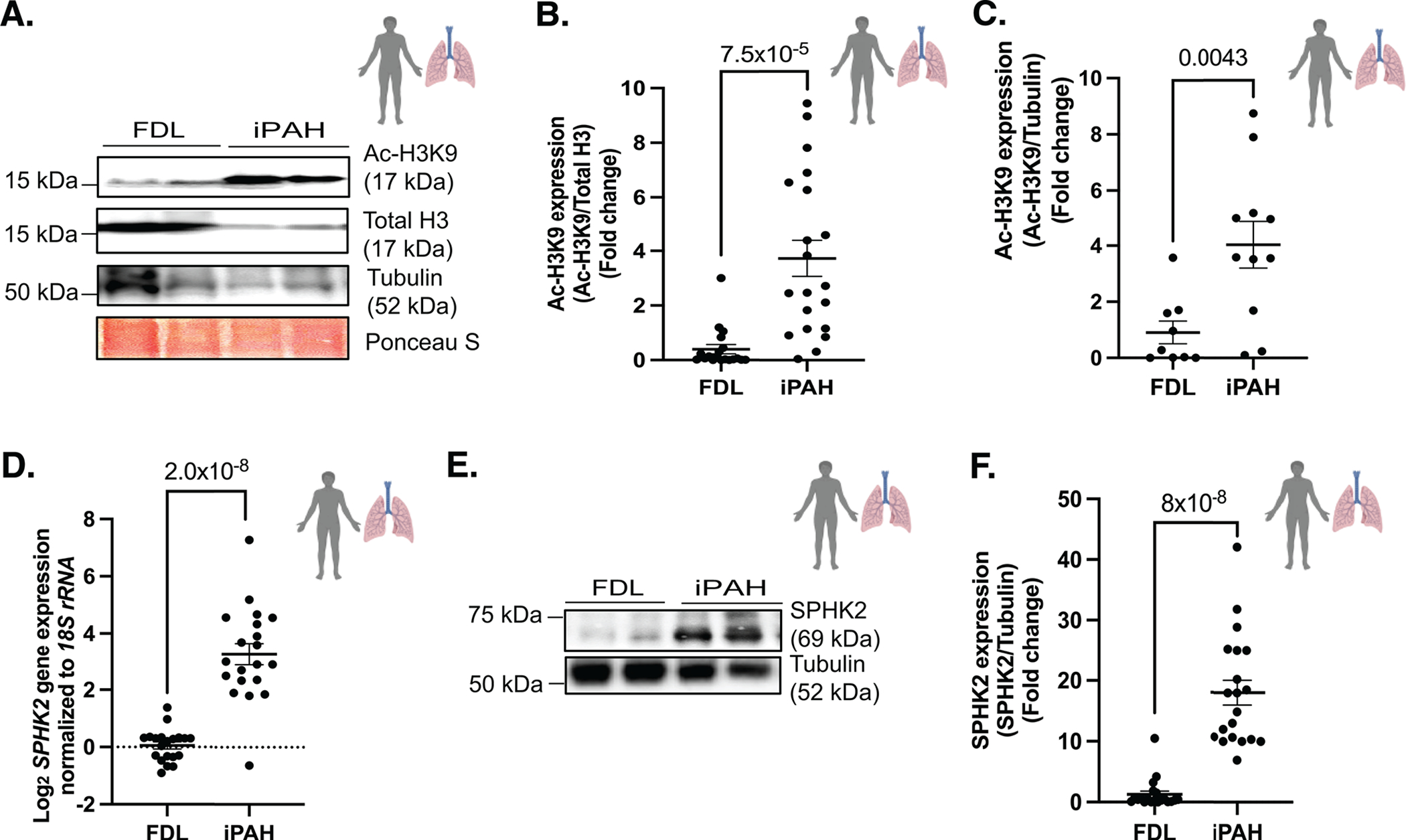
Figure 1. H3K9 acetylation and SPHK2 expression show a potential correlation in PAH patients’ lungs.

(A) Representative immunoblot probed for Ac-H3K9, total H3, tubulin and Ponceau S staining in protein lysates of human idiopathic pulmonary arterial hypertension (iPAH: type of Group 1 PH) lung or failed donor lung (FDL) tissue specimens and (B) quantitation of Ac-H3K9/Total H3, n=19–20 (C) quantitation of Ac-H3K9/Tubulin in protein lysates of human iPAH (n=11) or FDL (n=9). (D) SPHK2 expression levels normalized against 18S rRNA in iPAH lung and FDL tissues. n=20 (E) Representative immunoblot probed for SPHK2 and Tubulin in protein lysates of human iPAH (type of Group 1 PH) lung or FDL tissue specimens and (F) quantitation of SPHK2/Tubulin in protein lysates of human iPAH lung or FDL, n=20. P values are calculated using unpaired t-test and results are shown as means ± SEM.
