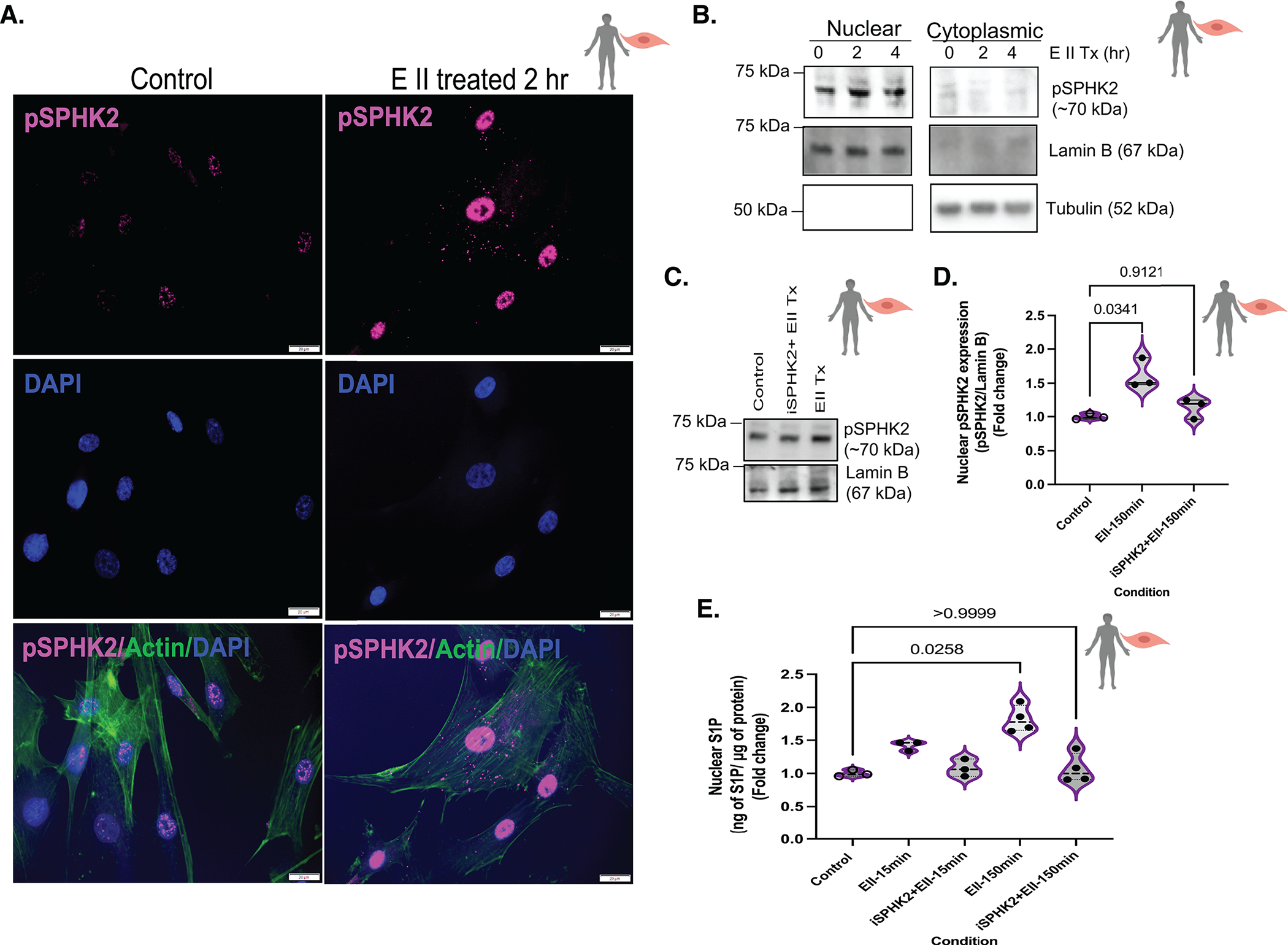
Figure 4. EMAP II promotes nuclear activation of SPHK2 that in turn generates nuclear lipid, S1P in vascular SMCs.

(A) Representative immunocytochemistry images of pSPHK2 (pink), actin (green, cytoplasmic marker) and DAPI (blue, nuclear) coimmunostaining in EMAP II treated (2 hr) or vehicle treated fixed hPASMCs, scale bar is 20 μm, n=3. (B) Representative immunoblot probed for pSPHK2, tubulin and lamin B in cytoplasmic and nuclear fractions of hPASMCs following EMAP II treatment for 0, 2 and 4 hours, n=3. (C) Representative immunoblot probed for pSPHK2 and lamin B in nuclear fractions of hPASMCs following EMAP II treatment (150 minutes) with or without SPHK2 inhibitor (D) quantification of nuclear pSPHK2/lamin B, n=3. (E) ELISA-nuclear C18-S1P levels normalized against 1 μg of nuclear proteins in the nuclear fractions of hPASMCs following EMAP II for 15 or 150 minutes with or without SPHK2 inhibitor, n=3 or 4/group. P values are calculated using Kruskal-Wallis against control or Kolmogorov-Smirnov non-parametric test and results are shown as median and inter-quartile range.
