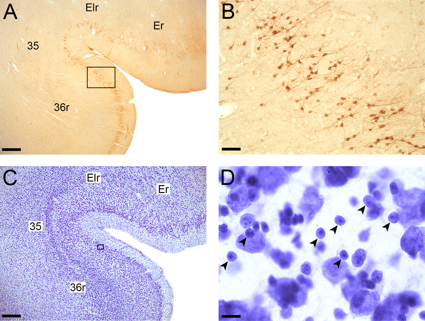
Figure 2.

Examples of Bcl2 and Nissl-stained immature neurons in the perirhinal cortex. A. Bcl2-stained section showing areas Er and Elr of the entorhinal cortex and areas 35 and 36r of the perirhinal cortex; Magnification: 2X; Scale bar: 500 µm. B. Bcl2+ cells and thin processes visible within layer II of the perirhinal cortex; 20X; Scale bar: 50 µm. C. Nissl-stained section showing areas Er and Elr of the entorhinal cortex and areas 35 and 36r of the perirhinal cortex; 2X; Scale bar: 500 µm B. Nissl-stained immature neurons (small arrowheads) within layer II of the perirhinal cortex; 100X; Scale bar: 10 µm.
