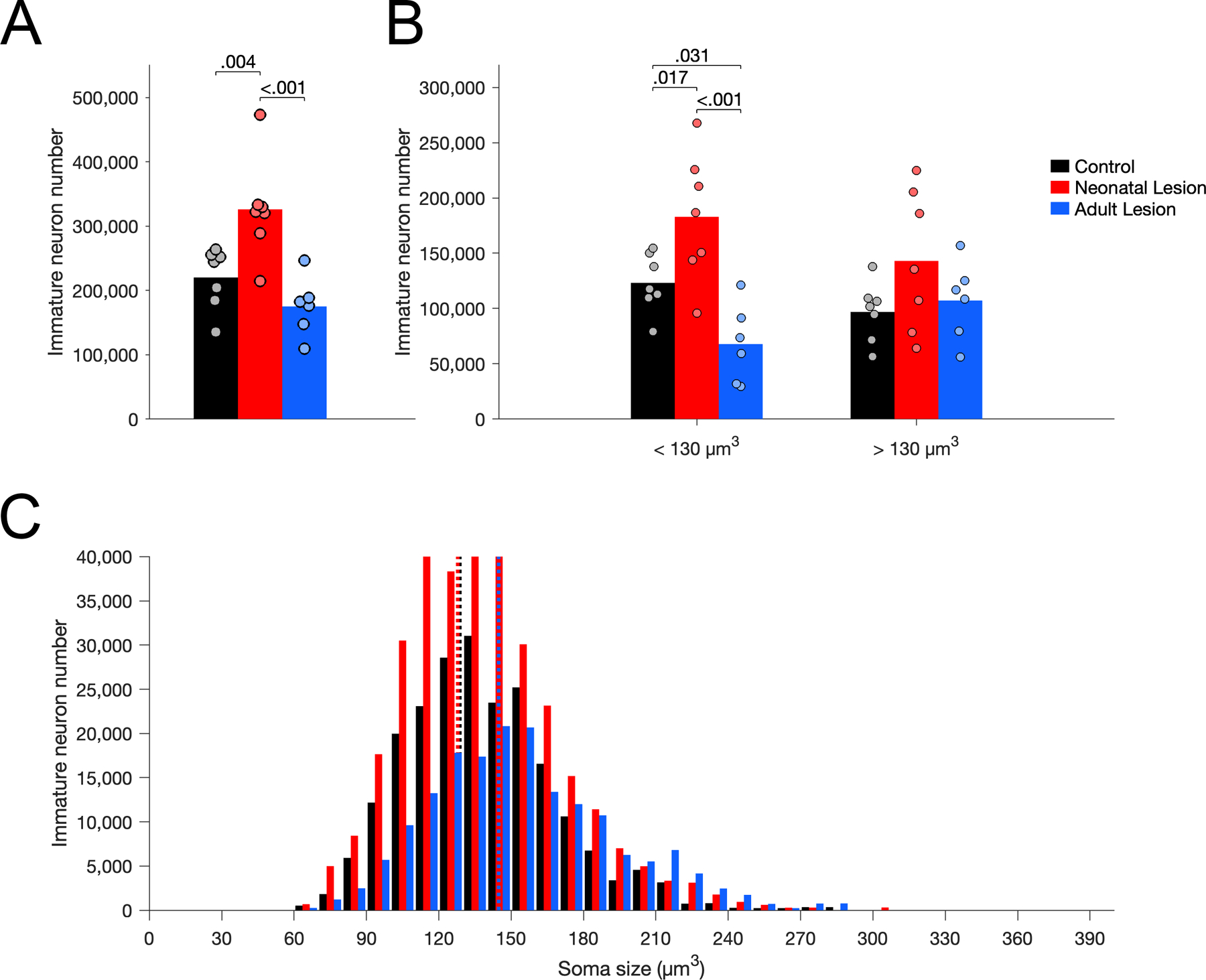
Figure 8.

Number of Nissl-stained immature neurons in layer II of area 36 in unoperated control (black), neonatal hippocampal-lesioned (red) and adult hippocampal-lesioned (blue) monkeys. A. Total number of immature neurons. B. Number of small and large immature neurons, below and above the median soma size of controls (130 µm3). C. Distribution of immature neuron soma size (in cubic micrometers, µm3). Dotted lines represent the average soma size for each group (controls: 129 µm3; neonatal lesion: 128 µm3; adult lesion: 145 µm3).
