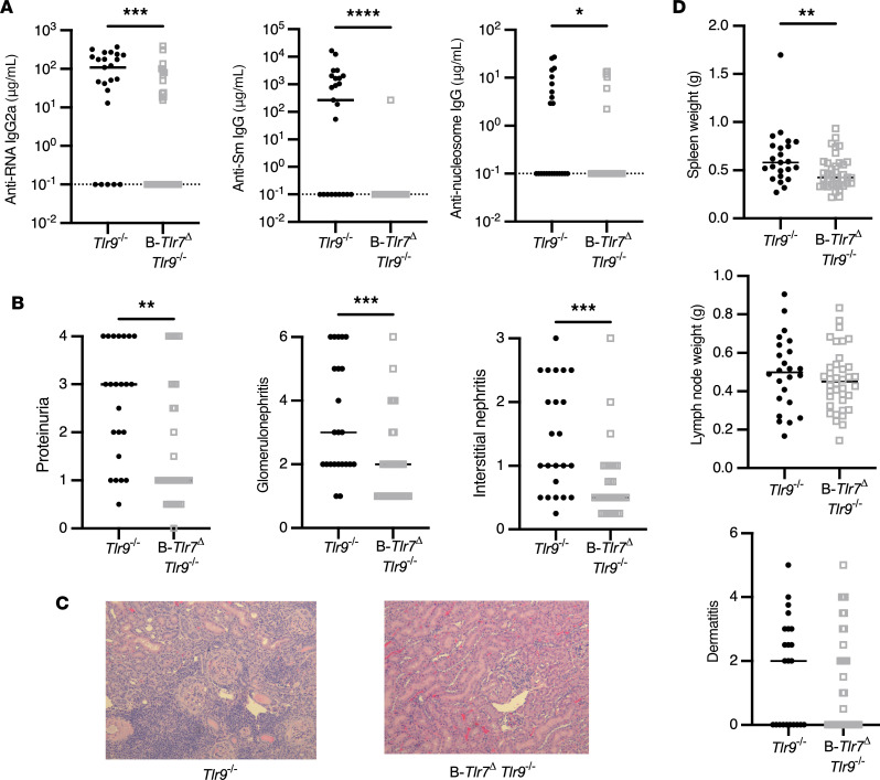
Figure 5. B cell–intrinsic TLR7 drives severe renal disease, splenomegaly, and the anti-RNA–associated autoantibody responses in TLR9-deficient mice.
Control (Tlr7fl/fl Tlr9–/– and Tlr7fl/y TLR9–/–) and B-Tlr7Δ Tlr9–/– (CD19-Cre+/– Tlr7fl/fl Tlr9–/– and CD19-Cre+/– Tlr7fl/y Tlr9–/–) mice were aged until 16 weeks (female) or 19 weeks (male). (A) Serum concentrations of anti–RNA IgG2a, anti–Sm IgG, and anti–nucleosome IgG in control and B-Tlr7Δ Tlr9–/– (n = 23 and n = 37, respectively). (B) Evaluation of renal disease including proteinuria, glomerulonephritis, and interstitial and perivascular infiltrates in control versus B-Tlr7Δ Tlr9–/– mice (n = 23 and n = 37, respectively). (C) Representative images of H&E-stained kidney sections for indicated genotypes. Original magnification, 200×. (D) Quantification of spleen weight, lymph node weight, and dermatitis in control versus B-Tlr7Δ Tlr9–/– mice (n = 23 and n = 37, respectively). Scatterplots display data from individual mice, with black lines showing median values and dotted lines indicating the lower limit of detection. *P < 0.05, **P < 0.01, ***P < 0.001, ****P < 0.0001 by 1-tailed Mann-Whitney U test.

