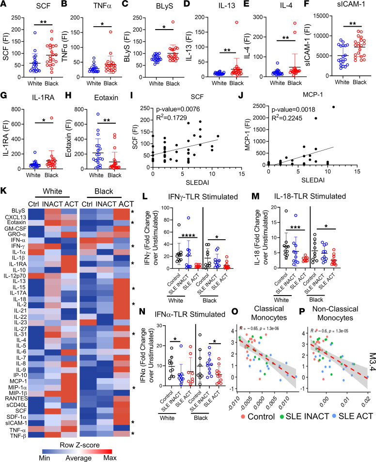
Figure 8. Pro-inflammatory cytokines vary in patients with SLE by ancestry and shape the TLR immune response.
Pro-inflammatory soluble mediators were measured by multiplex or ELISA. Significant cytokine differences between Black (n = 21) and White (n = 19) SLE patients included increased (A) SCF, (B) TNF-α, (C) BLyS, (D) IL-13, (E) IL-4, (F) sICAM-1, and (G) IL-1RA in Black patients and increased (H) eotaxin in White patients. Linear regression analyses show (I) SCF and (J) MCP-1 to increase with SLE disease activity (SLEDAI) (n = 40). (K) A heatmap summary of the MFI supernatant levels following 24-hour whole blood stimulation with TLR4/7/8/9 agonists for each disease group is shown. Soluble mediator levels are displayed on a color scale ranging from blue (protein levels below the mean) to red (protein levels greater than the mean) using a column z score. Significant differences between Black and White disease groups are noted. The most significant fold-change differences over unstimulated culture samples were in the IFN pathways, including (L) IFN-γ, (M) IL-18, and (N) IFN-α. TLR-stimulated culture supernatant levels of IFN-γ negatively associated with mean ISG gene expression modules in (O) classical and (P) nonclassical monocytes by linear regression analysis. Statistical significance was determined using a Mann-Whitney U test (P < 0.05), and all FDR q values were used for multiple comparisons. Mean ± SD is shown. *P < 0.05, **P < 0.01, ***P < 0.001, ****P < 0.0001. FI, fluorescence intensity.

