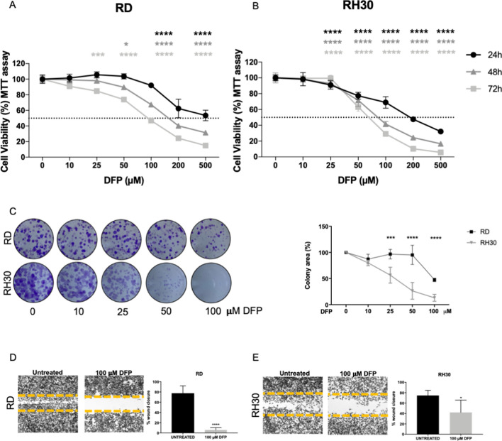
Fig. 4.
Iron chelation affected RMS cell viability, clonogenic and wound repair capacity. MTT assay after treatment with 10–25–50–100–200–500 µM of Deferiprone (DFP) for 24–48–72 h in RD (A) and RH30 (B) cells, representing human ERMS and ARMS, respectively (N = 3). C Clonogenic assay in RD and RH30 after treatment with 10–25–50–100 µM DFP for 48 h followed by 5 days without DFP. The representative images showed the formed colonies stained with crystal violet and the correspondent graphs represent the colorimetric quantification of the solubilized crystal violet (N = 3). Wound healing assay in RD (D) and RH30 (E) after treatment with or without 100 µM DFP for 16 h for RD and for 10 h for RH30. (N = 3). Statistic was obtained by two-way anova in A and B and Students’ t test for unpaired data in C, D, E. The differences were considered as significant for: ****P < 0.0001, ***P < 0.001, **P < 0.01

