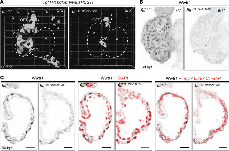
Figure 7. Aberrant activation of the Notch and Hippo signaling pathways in Flii-deficient ventricles.
(A) Representative 3D volume renderings of TP1bglob:VenusPEST of flii+/? sibling and fliiD110fs/D110fs hearts at 96 hpf. White dotted line outlines the heart. Note that there is no Notch reporter expression in the ventricle of fliiD110fs/D110fs hearts, whereas there is expression in their AVC and OFT. Each group n = 5. V, ventricle; A, atrium; AVC, atrioventricular canal; OFT, outflow tract. (B) Representative maximum-intensity projections of wholemount flii+/? sibling and fliiD110fs/D110fs hearts at 60 hpf stained for Wwtr1. flii+/? siblings n = 7, fliiD110fs/D110fs n = 11. (C) Corresponding confocal sagittal sections of the wholemount ventricles shown in B. Nuclei are counterstained with DAPI, and cardiomyocyte F-actin myofibrils are marked with myl7:LIFEACT-GFP expression; scale bars, 20 μm.

