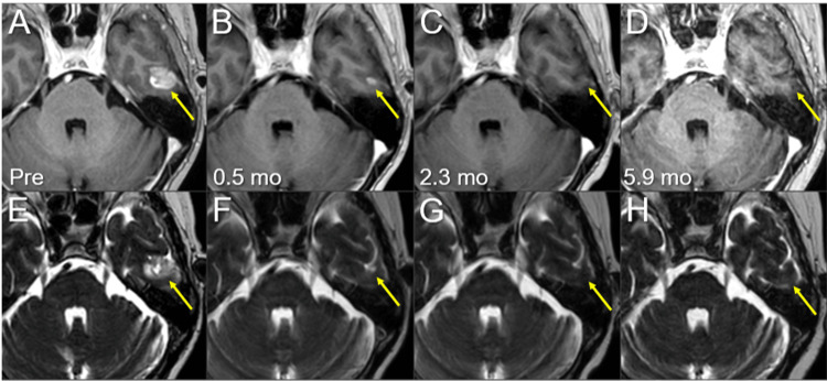
Figure 8. Magnetic resonance images before and after the third SRS of the left temporal new lesion.
The images show (A-D) axial CE-T1-WIs and (E-H) axial T2-WIs (A, E) five days before (Pre) the third SRS (17.3 mos after the initiation of CRT); (B, F) at 0.5 mo after the initiation of the third SRS; (C, G) at 2.3 mos; and (D, H) at 5.9 mos (23.9 mos after CRT).
(A-H) These images are shown at the same magnification and coordinates under co-registration and fusions. (A-H) The solid enhancing lesion (arrows in A and E) shrank remarkably at 0.5 mo (arrows in B and F) and disappeared at 2.3 mos (arrows in C and G). The lesion remained completely regressed without adverse radiation effects at 5.9 mos (arrows in D and H).
CE, contrast-enhanced; WIs, weighted images; SRS, stereotactic radiosurgery; CRT, chemoradiotherapy; mo, month

