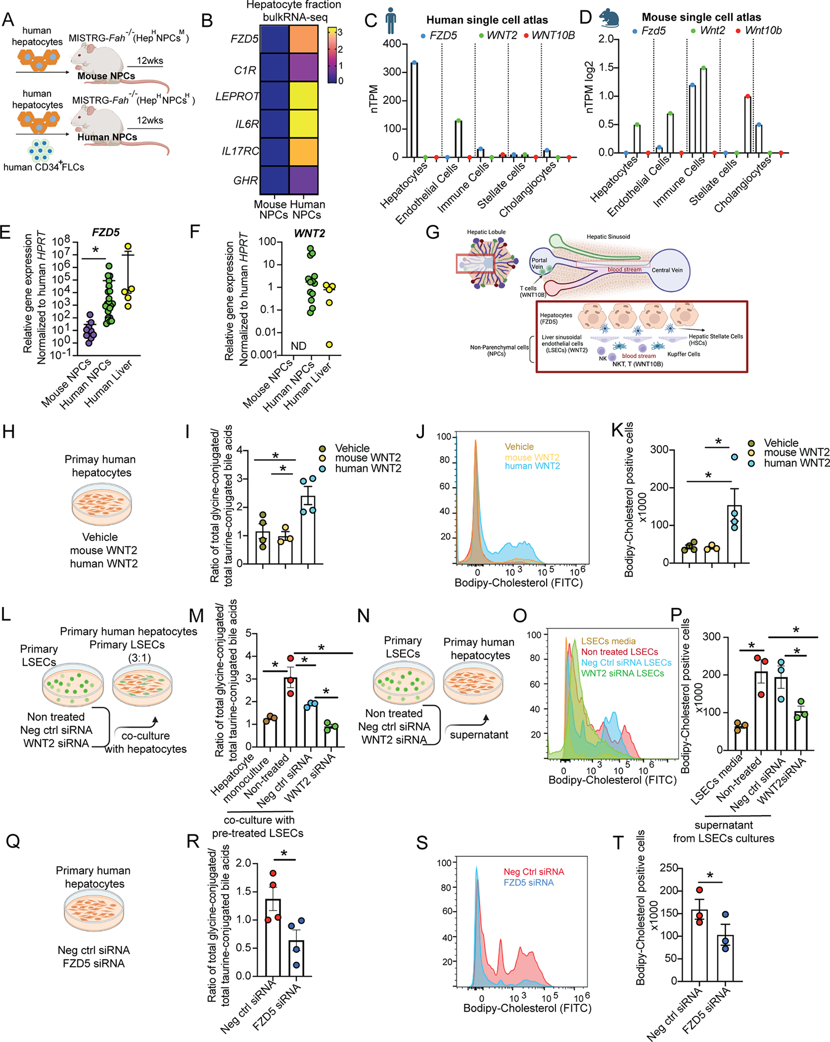
Figure 5. Endothelial cell-derived WNT2 regulates the metabolic function of human hepatocytes through FZD5.

(A) Adult MISTRG-Fah−/− mice were engrafted with human hepatocytes and human CD34+ FLCs (human NPC group) or with only human hepatocytes (mouse NPC group).
(B) Bulk RNA-seq in human hepatocytes. Average fold change in the expression of receptor genes in mice with human NPCs relative to mice with mouse NPCs.
(C,D) Gene expression of human/mouse FZD5 and their ligands across different cell types in human and mouse liver atlas datasets1. See also Figure S7.
(E, F) Human FZD5 and WNT2 gene expression by RT-qPCR in the hepatocyte fraction from MISTRG-Fah−/− mice and in healthy human liver tissue from partial hepatectomies.
(G) Schematic representation of liver cell location. Cartoon was made using BioRender.
(H-K) Primary human hepatocytes treated with human WNT2, mouse Wnt2 or vehicle (DMSO) for 24 hours.
(L-P) Co-culture for 24 hours of primary human hepatocytes with primary human LSESs directly in the same plate or indirectly after supernatant transfer from LSECs. Primary human LSECs were pre-treated before co-culture for 24hours with negative control (Neg ctrl) siRNA or WNT2 siRNA or left untreated.
(Q-T) Primary human hepatocytes treated with siRNA silencing human FZD5 or its negative control (neg ctrl) for 24 hours.
(H-T) Primary bile acids were measured by HPLC-MS/MS in hepatocytes. Hepatocytes were treated with labeled cholesterol (Bodipy) for two hours and analyzed by flow cytometry.
Each dot in the graphs is a biological replicate from at least 2 independent experiments. Data represent mean ± SEM. *p <0.05. See also Figure S8.
