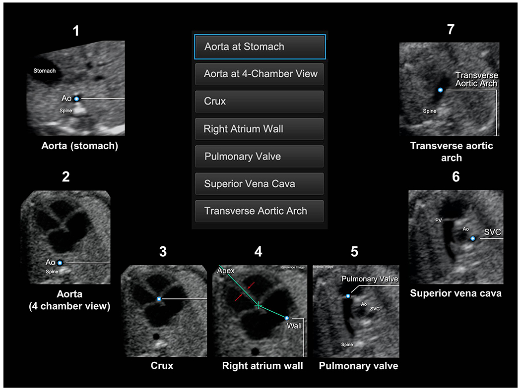
Figure 1: Seven anatomical structures within the heart are marked using the Anatomic Box® tool.

(also see Videoclip S1): 1) cross-section of the aorta at the level of the stomach; 2) cross-section of the aorta at the level of the four-chamber view; 3) crux; 4) right atrial wall; 5) pulmonary valve; 6) cross-section of the superior vena cava; and 7) transverse aortic arch. Ao, aorta; PV, pulmonary valve; SVC, superior vena cava.
