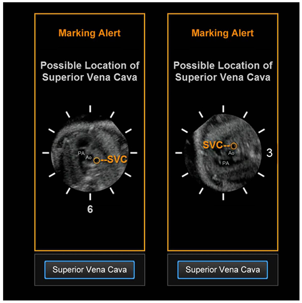
Figure 3: Superior vena cava alert.

This type of marking alert notifies the user that the superior vena cava may be in a different location that what is expected. This is because the fetal spine in the STIC volume dataset being analyzed by FINE is at 3 o’clock. Thus, there may be difficulty in marking the cross-section of the superior vena cava using Anatomic Box®. This alert includes a reference movie that will automatically play and depicts the orientation/position of the pulmonary artery (PA), aorta (Ao), and superior vena cava (SVC) when the spine is at 6 o’clock (left image) and 3 o’clock (right image). The movie is intended to help the user recognize that for their STIC volume, the superior vena cava may be in a location similar to that depicted in the movie.
