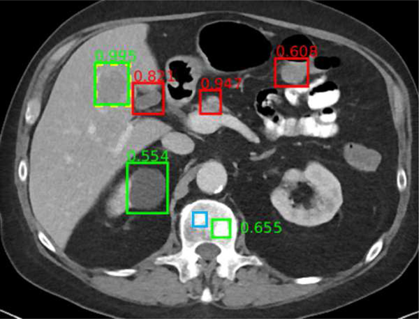
Fig. 6.
Example universal lesion detector for abdominal CT. In this axial image through the upper abdomen, a liver lesion was correctly detected with high confidence (0.995). A renal cyst (0.554) and a bone metastasis (0.655) were also detected correctly. False positives include normal pancreas (0.947), gallbladder (0.821), and bowel (0.608). A subtle bone metastasis (blue box) was missed. Reproduced from [186].

