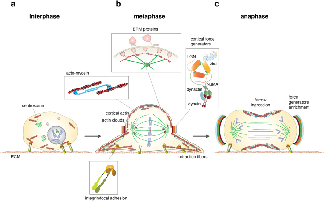
Figure 1. Morphological changes instructing spindle orientation during mitotic progression.
a) Distribution of integrin/focal adhesion adhesive cues, F-actin cytoskeleton and chromosomes in interphase and prometaphase. Centrosomes are positioned near the nucleus, NuMA is nuclear, and ERM (Ezrin-Radixin-Moesin) proteins are found in a closed inhibited conformation. b) Rounded-up morphology of cells in metaphase, with sister chromatids congressed on the metaphase plate and a bipolar spindle. The spherical shape is sustained by an actomyosin layer (1, which is connected to the plasma membrane near actin clouds by activated ERM proteins (2). Mitotic cells maintain contact to the substrate via mitotic β1-integrin containing focal adhesion complexes anchored to actin-based retraction fibres (3). The mitotic spindle axis is maintained aligned to the substratum by dynein–dynactin cortical force generators recruited to cortical crescent above the spindle poles by Gαi–LGN–NuMA complexes (4). Actin clouds formed by F-actin localizing near the spindle poles contribute to spindle orientation as well. c) At anaphase, force generators level increases symmetrically above the spindle poles. At this point, NuMA is anchored to the membrane independently of LGN, by binding to membrane phospholipids, after its phosphorylation by CDK1 is relieved (not shown). This allows generation of stronger dynein-based traction forces that are needed to separate sister chromatids in synergy with pushing and pulling forces acting at the kinetochore level. Anaphase is also associated with actomyosin cortex reorganization, whereby actomyosin enriches at the equatorial region of the cell to promote cytokinetic cleavage furrow ingression.

