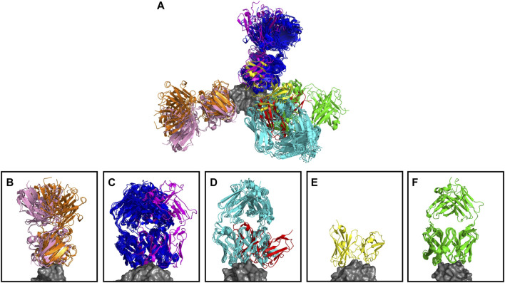
FIGURE 4.
Anti-lysozyme antibodies. Crystal structures of 53 antibody–lysozyme complexes are shown aligned on the antigen structure (gray). Antibodies are colored according to the clusters assigned by SPACE2. (A) Overlay showing all 53 antibody–lysozyme complexes. (B–F) Each panel shows all antibodies that bind to one of the five lysozyme epitopes as defined by Ab-ligity (Wong et al., 2021). Panels (B–D) Each contain two sets of antibodies that do not overlay perfectly indicating a difference in binding pose. SPACE2 separates antibodies binding to the same epitope in a different binding pose into distinct clusters as indicated by coloring.

