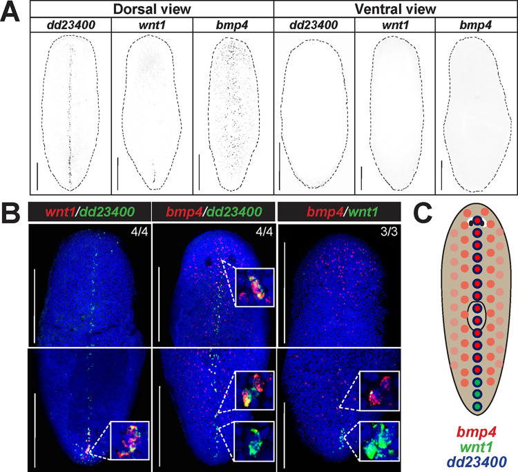
Fig 1. bmp4 and wnt1 co-express with dd23400+ dorsal midline cells in a regionally distinct manner.
(A) Fluorescent in situ hybridization (FISH) detecting dd23400 on the dorsal midline, wnt1 on the dorsal posterior midline, and bmp4 in a dorsal midline-centered gradient with reduced expression in the far posterior. Dotted line indicates animal outline. Scale bars represent 150 μm with dorsal or ventral views indicated. (B) Double FISH detecting co-expression of wnt1, dd23400, and bmp4. Left: 88.9% of the cells that were wnt1+ were also dd23400+ (64/72 cells from 4 animals). Middle: 55.4% of dd23400+ cells also expressed bmp4 (169/305 cells from 4 animals). Right: Only 12.1% of cells expressing wnt1+ also co-expressed bmp4 (4/33 cells from 3 animals, top inset). The wnt1+ cells that expressed bmp4 were consistently the anterior-most cells of the wnt1 domain. By contrast, 87.9% (29/33 wnt1+ cells from 3 animals, bottom inset), all located at the posterior tip, did not co-express bmp4. (C) Schematic illustrating separation of bmp4 and wnt1 domains on the dorsal midline. Along dd23400+ dorsal midline muscle cells, wnt1 and bmp4 expression defines largely nonoverlapping AP domains within posterior. Within the wnt1+ domain of the representative image, wnt1+ cells in the anterior co-expressed bmp4 whereas cells in the posterior of the domain lacked bmp4 co-expression. Scale bars represent 150 μm.

