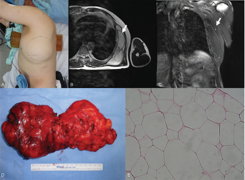
Figure 1.

A 60-year-old male admitted with a swollen left chest wall without any symptom in 2015 (A). Based on the the magnetic resonance imaging (MRI) findings, an approximately 159 x 144 x 38 mm septate mass lying beneath serratus anterior muscle was identified in the left chest wall (B, C). A complete surgical excision was performed (D). The submitted specimen is an ovoid mass, measuring 160 x 85 x 75 mm with a pale-brown and a thin fibrotic capsule. In the pathological findings (x 400), the neoplasm was composed of mature white adipose tissue with few adipocytes suggesting a benign lipoma (E).
