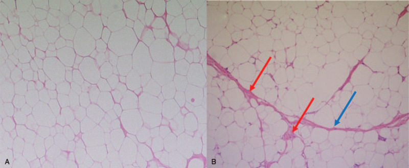
Figure 5.

Pathologic finding (x 100) of excised tissue in 2015 shows the mature adipose tissue with few adipocytes suggesting a benign lipoma (A). Pathologic findings (x 100) of the excised tissue in 2019 show multiple septa (blue arrow) and variation in cell size, and focal adipocytic nuclear hyperchromasia (red arrow) suggesting WDLS (B).
