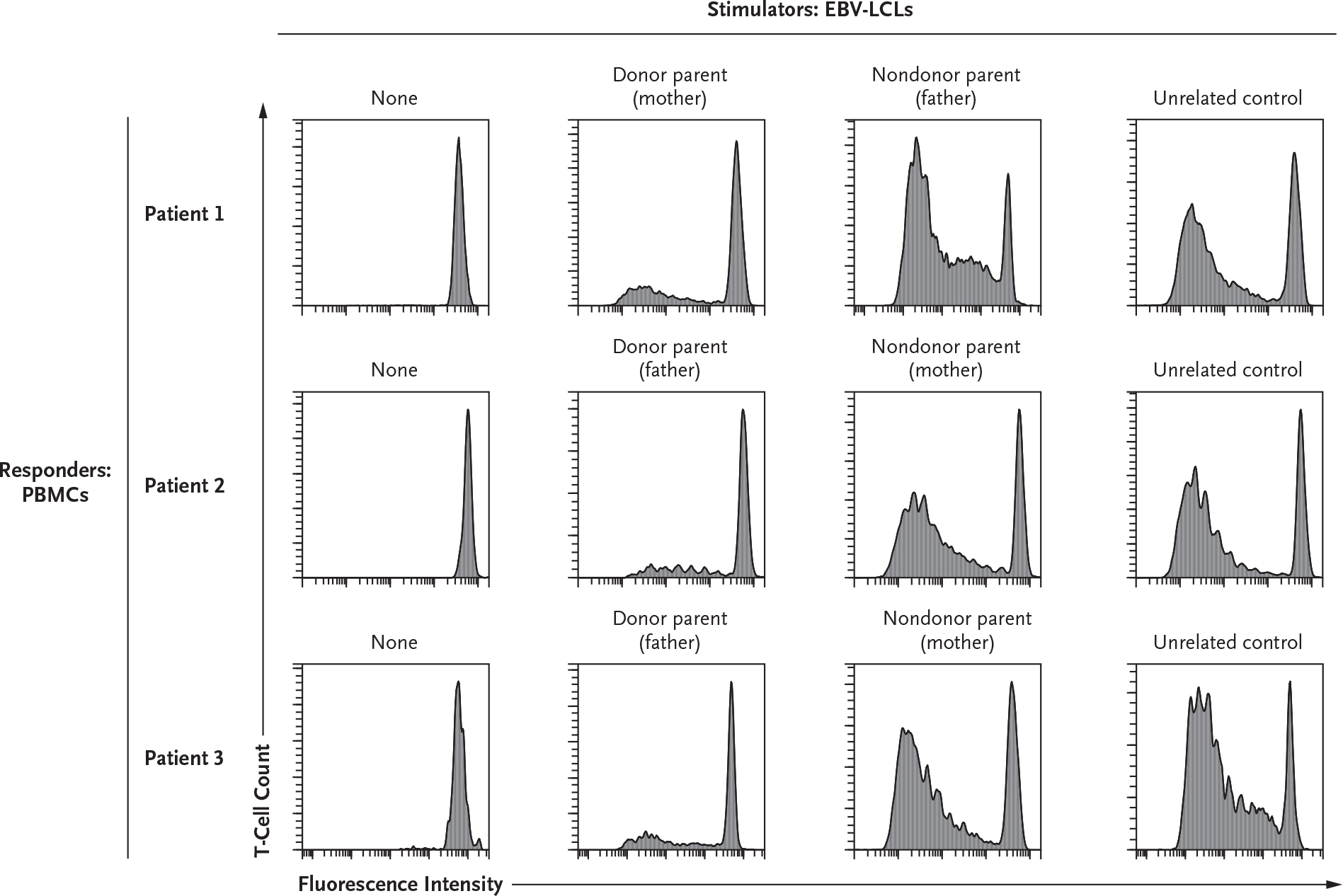
Figure 2. One-Way Mixed-Lymphocyte Cellular Assay of T-Cell Alloreactivity at 1 Year after Kidney Transplantation.

Peripheral blood mononuclear cells (PBMCs), which were used as responder cells, were isolated from the three patients at least 1 year after their kidney transplantation (KT). The PBMCs were stained with CellTrace Violet (Invitrogen) before coculture with stimulator Epstein–Barr virus (EBV)–transduced B lymphoblastoid cell lines (EBV-LCLs) previously generated from the parents of the patients or from an unrelated healthy third-party control. CellTrace Violet fluorescence intensity (on a logarithmic scale) for CD3+CD19− cells (x axis) is shown plotted against T-cell count (y axis); a loss of fluorescence indicates cell proliferation. T-cell proliferation was measured after 6 days of culture and obtained at a responder-to-stimulator ratio of 2:1. T cells of both of the two siblings (Patients 1 and 2) and of Patient 3 showed functional tolerance to EBV-LCLs derived from their respective donors, whereas they were immune competent and proliferated in the presence of EBV-LCLs derived from nondonor parents or a healthy, unrelated control. The plots in the first column show the absence of T-cell proliferation when PBMCs were cultured in medium alone. The results shown are representative of three replicates for each combination of stimulator and responder cells.
