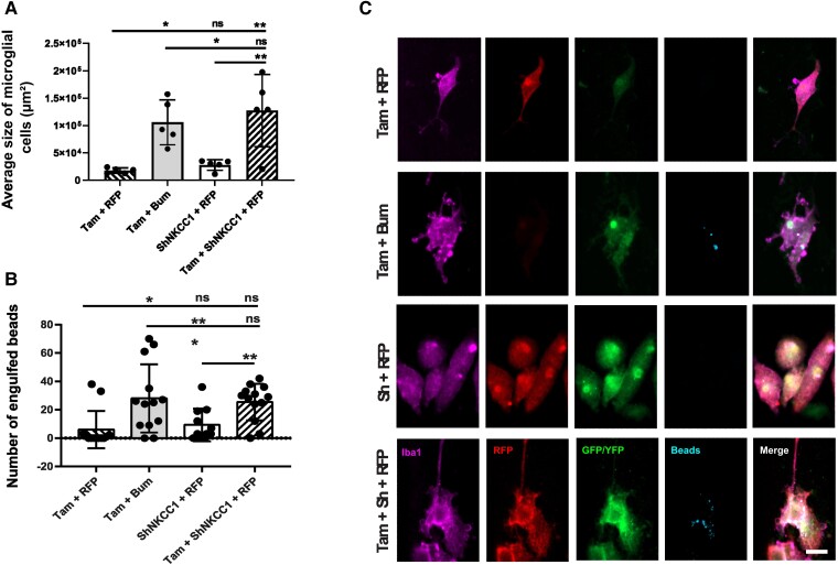
Figure 3.
Effect of impaired chloride uptake in microglia morphology and phagocytosis capacity. (A) Average size of primary microglia cells from CX3CR1-Cre ERT2 mice under different conditions: co-transfected with pSico-sh Nkcc1 and pCAG-RFP, treated with bumetanide (Bum) and tamoxifen (Tam), transfected with pCAG-RFP and treated with Tam, co-transfected with pSico-sh Nkcc1 and pCAG-RFP and treated with Tam. n = 4 wells per condition and five cells chosen randomly per well. (B) Number of latex beads engulfed by cells in the different conditions. n = 4 wells per condition and 12 cells chosen randomly per well. (C) Immunostainings of the primary microglia cell culture from CX3CR1-Cre ERT2 mice under the different conditions. Anti-Iba1 was used to label microglial cells (purple), anti-RFP to label the pCAG-RFP plasmid (red) and GFP to enhance the pSico-sh Nkcc1 fluorescence (green). YFP was expressed when cells were treated with Tam (green) and the beads were labelled with a Rabbit IgG-DyLight 633 Complex (blue). All data were analysed using a one-way ANOVA with Dunnett's post hoc test. *P < 0.05; **P < 0.01; ***P < 0.001; ns = not significant.

