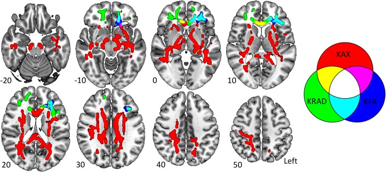
Figure 1.
Diffusion kurtosis imaging regions of interest. Regions of interest (ROIs) from the skeletonized data have been inflated using tbss_fill for visualization. The Axial kurtosis (KAX) ROI, the kurtosis-fractional anisotropy (KFA) ROI, the radial kurtosis (KRAD) ROI, and areas of overlap between ROIs are displayed. Z-coordinates are displayed for each slice. Images are displayed using radiological convention. The figure was made using MRIcroGL.

