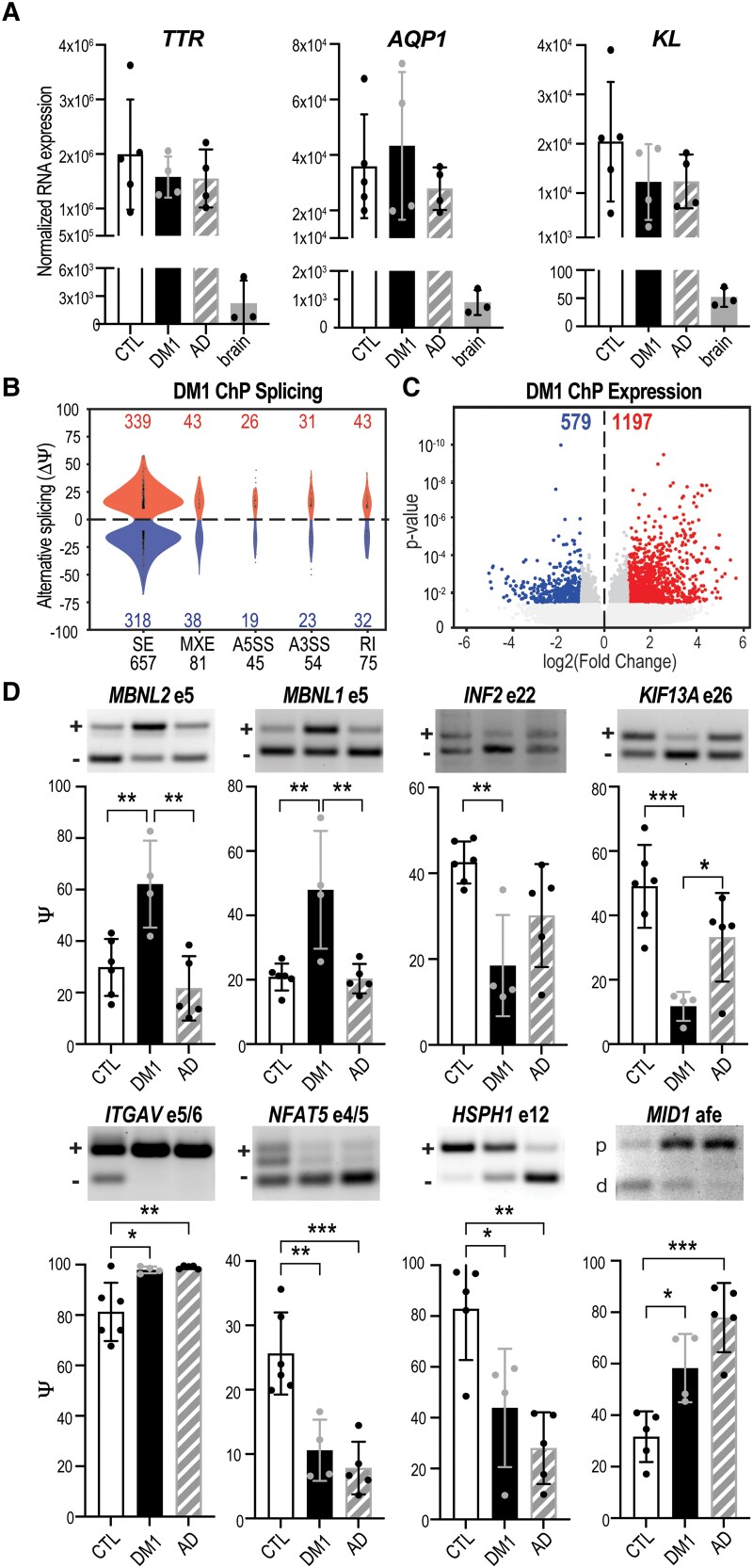
Figure 4.
ChP spliceopathy in DM1. (A) Normalized RNA expression (RNA-seq) for choroid plexus (ChP) markers TTR, AQP1 and KL for neurologically unaffected controls (CTL), myotonic dystrophy type 1 (DM1) and Alzheimer’s disease (AD), together with non-ChP brain tissues (brain). (B) Violin plots of AS events (Ψ) quantified by change in per cent spliced in (ΔPSI). A5SS = alternative 5′ splice site; A3SS = alternative 3′ splice site; MXE = mutually exclusive exon; SE = skipped exon. (C) DE of RNA transcripts showing the log2 transformed fold change plotted against significance (P-value). (D) RT-PCR validations of splicing changes in DM1 ChP compared to CTL and AD for MBNL2 exon 5, MBNL1 exon 5, INF2 exon 22, KIF13A exon 26, ITGAV exons 5 and 6, NFAT5 exons 4 and 5, HSPH1 exon 12 and MID1 alternative first exon (afe). *P < 0.05, **P < 0.01, ***P < 0.001, one-way ANOVA.

