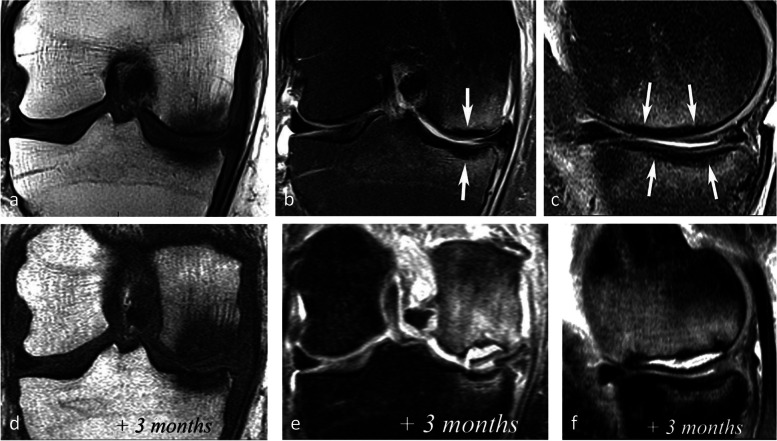
Fig. 17.
Negative prognostic value of low signal bone marrow on T2-weighted MR image. a The subchondral bone of the two medial femorotibial joint surfaces shows marked low signal intensity on a T1-weighted MR image. b, c Coronal and sagittal fat-suppressed T2-weighted MR images (b, c) show relatively wide and thick subchondral areas adjoining the articular surface, with low signal intensity (arrows), suggestive of SIF-ON. d–f Three months later, a significant bone marrow low signal intensity persists on T1-weighted image (d) and coronal and sagittal fat-suppressed T2-weighted images show pejorative evolution with a collapse of the tibial plateau and a wide subchondral bone dissection and chondral fracture in the lower pole of the condyle (e, f)

