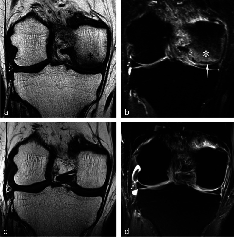
Fig. 19.

Borderline thickness of bone marrow low signal on T2-weighted image. a, b MRI shows low signal intensity on T1-weighted (a) and high signal intensity on fat-suppressed T2-weighted images in the medial femoral condyle (b) (asterisks). Immediately near the subchondral surface, fat-suppressed T2-weighted image shows a very thin layer of tissue of borderline thickness with low signal intensity (arrow in b). c, d In the present case, the follow-up at 3 months showed healing, with normalization of the signal in the inferior pole of the condyle
