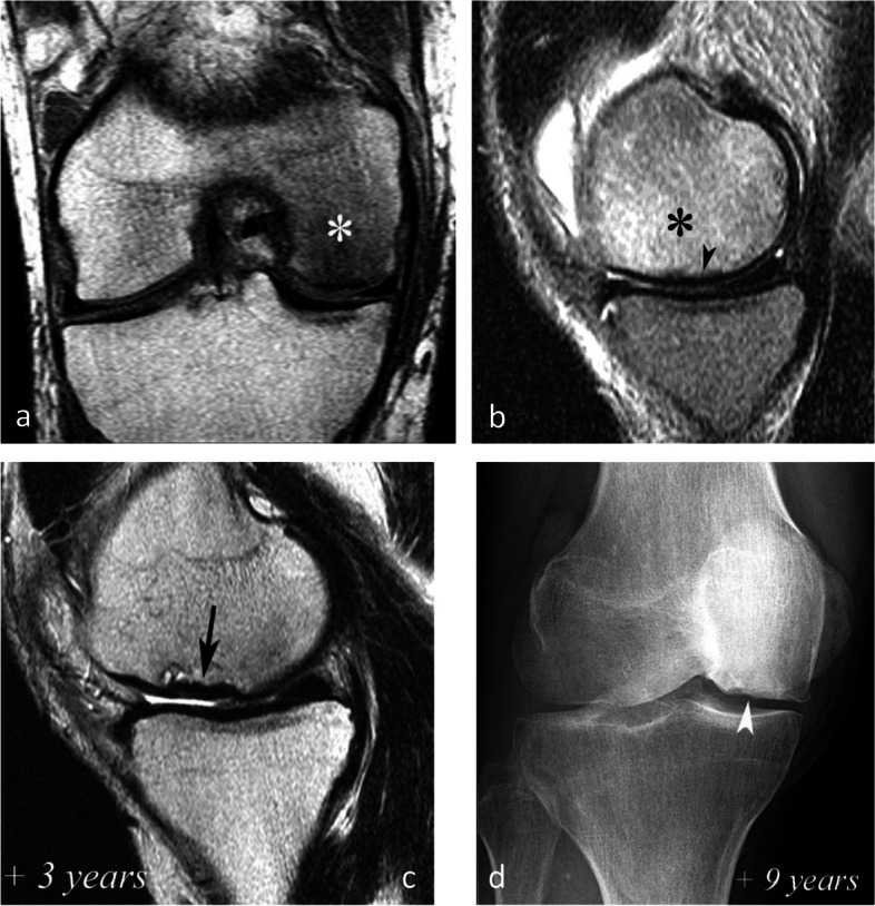
Fig. 20.

Evolution in two stages of subchondral insufficiency fracture (SIF). a, b Coronal T1-weighted (a) and sagittal T2-weighted (b) MR images show BME-like signal intensities in medial condyle (asterisks), except for a very thin (< 4 mm in thickness) subchondral low signal intensity area on the T2-weighted image (arrowhead in b). c MRI follow-up three years later with T2-weighted image (c) shows a small deformation of the lower pole of the condyle with thickening of the subchondral low signal intensity area, now exceeding 4 mm in thickness (arrows), suggesting the evolution of the SIF towards osteonecrosis (SIF-ON). d Nine years after the initial examination, a radiograph confirms an irreversible lesion, with deformation of the epiphyseal surface (arrowhead), which is moderate in this case and clinically well tolerated
