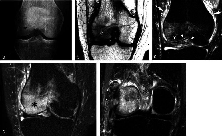
Fig. 24.
Typical pattern of complex regional pain syndrome type I (CRPS1). a Radiograph shows intense bone loss in the lower pole of a condyle (asterisk). b–e T1-weighted (b) and fat-suppressed T2-weighted (c–e) MR images show a BME-like pattern similar to that of a SIF (asterisks in b and c). Note the particularly intense edema-like signal intensity also in the soft tissues (arrows in c and asterisk in e), as well as a very fine linear area with intense high signal intensity, immediately adjacent to the subchondral bone plate (arrowheads in c)

