Fig. 25.

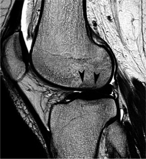
Trabecular fractures in case of CRPS1 (same MR examination as the previous figure). T2-weighted sagittal MR image shows thin, incomplete low-intensity linear images (arrows), as in a subchondral insufficiency fracture

Trabecular fractures in case of CRPS1 (same MR examination as the previous figure). T2-weighted sagittal MR image shows thin, incomplete low-intensity linear images (arrows), as in a subchondral insufficiency fracture