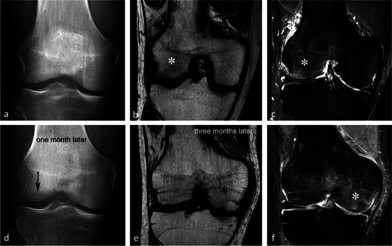
Fig. 26.
Migrating bone marrow edema (BME)-like lesion in CRPS1. a In early phase, the radiograph (a) was normal. b, c However, MR images showed typical BME-like signal appearing as low and high signal intensity on T1- (b) and fat-suppressed T2-weighted (c) images, respectively, in the lateral condyle (asterisks). d One month after the initial examination, radiograph show bone loss at the same location (arrow). e, f Three months after the initial examination, T1- and fat-suppressed T2-weighted images (e and f) show almost normal lateral condyle, while BME-like signal intensities have appeared in the medial condyle (asterisks)

