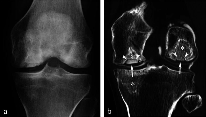
Fig. 31.

Radiographic of osteonecrosis of systemic origin. a Radiograph shows areas of heterogeneous bone sclerosis, in this case particularly prominent in the condyles. b CT arthrography shows areas of irregular bone densification corresponding to osteonecrotic lesions (asterisks) surrounded by sclerotic rims. Note collapsed segments in the weight-bearing areas of inferior condylar poles (arrows) associated with fractures within necrotic areas (arrowheads)
