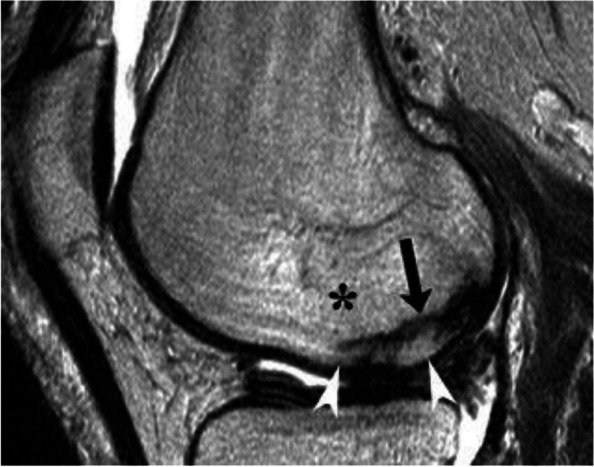
Fig. 4.

SIF in a patient with osteomalacia. T2-weighted MR image shows a low signal intensity band corresponding to a particularly thick trabecular fracture (arrow). The bone marrow of this portion of the condyle shows BME-like high signal intensity (asterisk). Note that the BME-like signal extends between the fracture and the bone-cartilage interface (arrowheads). That area of the subchondral bone is in continuity with the rest of the epiphyseal area
