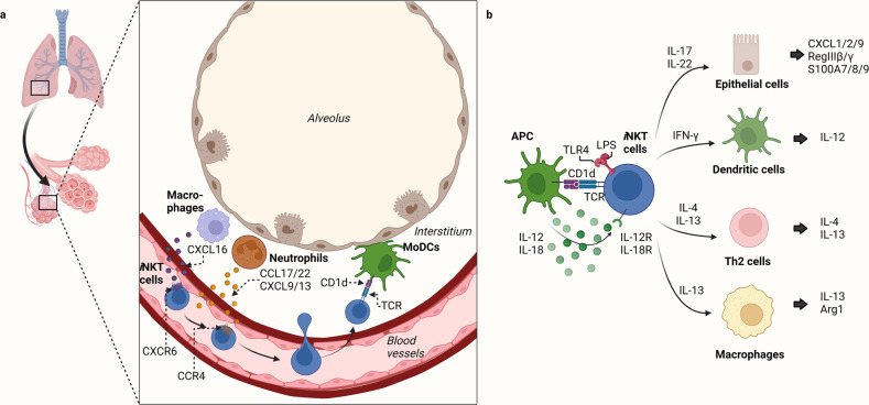
Fig. 1. Distribution of pulmonary iNKT cells and their interactions with other cells in the lung.
a Migration of iNKT cells to the lungs. After developing in the thymus, iNKT cells express CXCR6, a tissue localization molecule, and migrate to the CXCL16-expressing periphery. iNKT cells accumulate in the lumen of the lung microvasculature and then enter the lung tissue when neutrophils in the lung interstitium secrete the CCR4 ligands CCL17/22 and CXCL9/13. The neutrophils, therefore, guide the iNKT cells to the source of lung injury in the interstitium. The monocyte-derived DCs (moDCs) in this area present glycolipid antigens from the lung injury source to the iNKT cells, which become activated and then remain in the interstitium. b Crosstalk between iNKT cells and other cells in the lung. iNKT cells are activated by antigens expressed by lung antigen-presenting cells (APCs), such as MoDCs, by cytokines from other cells (such as IL-12/18), and by TLR ligands (e.g., LPS). The activated iNKT cells then secrete a variety of cytokines that regulate the function of many types of neighboring cells.

