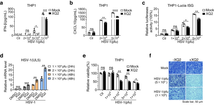
Fig. 5. XQ2 attenuates the host antiviral defenses.
a, b THP1 cells were pretreated for 3 h with DMSO or XQ2 (10 μM), and then stimulated by HSV-1 infection for 24 h. Induction of IFN-β (a) and CXCL10 (b) proteins was measured by ELISA. a p = 0.0264 (5 × 10⁴ pfu); p = 0.0001 (1 × 10⁵ pfu), b p = 0.0064 (2 × 10⁴ pfu); p < 0.0001 (5 × 10⁴ pfu). c THP1 luciferase reporter cells were exposed to DMSO or XQ2 (10 μM), and then stimulated by the indicated titer of HSV-1 infection for 24 h. Type I interferon response was indicated by luciferase activity. p = 0.043 (2 × 10⁴ pfu); p = 0.0018 (5 × 10⁴ pfu). d THP1 cells were pretreated for 3 h with DMSO or XQ2 (10 μM), and then stimulated by HSV-1 infection for indicated titer and times. Induction of HSV-1(UL5) mRNA was measured by qPCR. p = 0.0008 (1 × 10⁴ pfu, 24 h); p = 0.0007 (2 × 10⁴ pfu, 24 h); p = 0.0035 (1 × 10⁴ pfu, 48 h); p = 0.0029 (2 × 10⁴ pfu, 48 h). e THP1 cells were pretreated for 3 h with DMSO or XQ2 (10 μM), followed by the indicated titer of HSV-1 infection. The cell viability was measured by CCK-8 assay. p = 0.0024 (2 × 10⁴ pfu); p = 0.003 (5 × 10⁴ pfu); p = 0.0247 (1 × 10⁵ pfu). f L929 cells pretreated for 4 h with DMSO or XQ2 (10 μM), followed by HSV-1 infection. The proliferation of cells was examined by crystal violet staining. Data are representative of three independent experiments with similar results in (f), or three independent experiments in (a–e). (Data are presented as mean ± SD, n = 3 independent samples in (a–e), ns, not significant, *p < 0.05, **p < 0.01, ***p < 0.001, ****p < 0.0001 using one-way ANOVA with Dunnett’s post hoc test). Source data are provided as a Source Data file.

